A Step by Step Guide to Reviewing and Interpreting Pathology & Diseases Ultra-Widefield Retinal Imaging Recorded Webinar
When taking on new clinical diagnostic equipment it can be quite bewildering to view the eye in different ways. Often the new imaging techniques show structures in ways that look so different as to cause confusion.
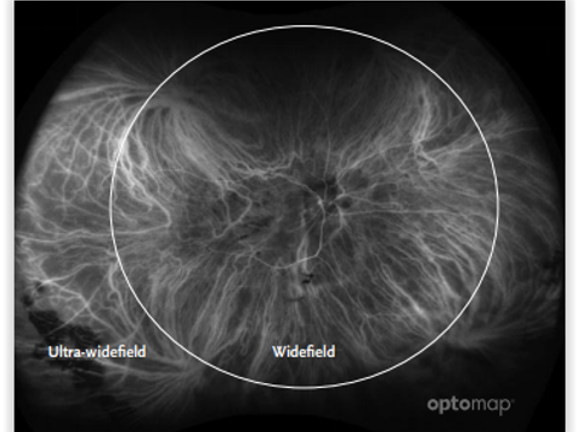
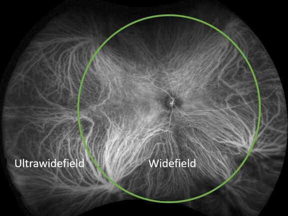
This webinar starts with a section on how to obtain the best possible images for review. There will then be a section on the anatomy of the eye and annotated images of ultra-widefield images are shown to demonstrate how much of the retina is seen and to understand where various retinal disorders are. Simon Browning sets out a step-by-step approach to reviewing images giving the reasons why things are done in that order and the outcomes of each step.
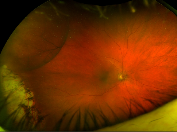
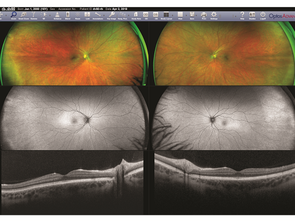
The webinar includes images from different instruments to demonstrate the logical process of the image review. At each stage the differential diagnoses are considered and the additional diagnostic tests that can be carried out to confirm or rule out potential diagnoses.
CPD Points: 1
CPDpoints.com credits: 1
Expiry Date: 31/12/2024
Sponsored by