New Technology, New Pathology Multi-modal imaging as an enhancement to standard retinal examinations
As optometrists continue to invest in new diagnostic technology such as OCT and ultra-widefield retinal imaging, many embark on a natural steep learning curve as they get to grips with new imaging techniques.
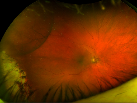
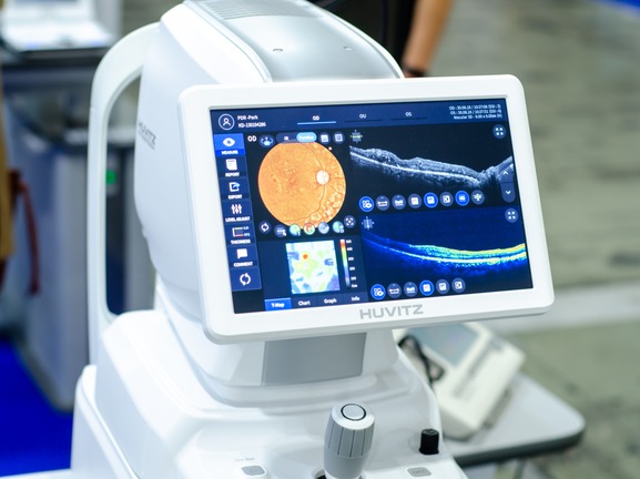
Case studies will focus around the key features of common ocular pathologies to help optometrists make an accurate diagnosis using ultra-widefield, autofluorescent and OCT imaging and associated diagnostic software. The diagnosis and management of cases of common ocular disease will be presented including:
- Naevus
- Melanoma
- Peripheral retinal disease: e.g. lattice degeneration
- Central retinal disease: e.g. ARMD, Diabetic retinopathy
The lecturer will also examine how to communicate effectively when referring a patient, using for example systems such as the MOLES diagnostic tool developed by Professor Bertil Damato and now adopted as a national guideline by LOCSU, so that the person triaging the referral is able to make the best decision in sending the patient to the most appropriate setting for their consultation.
This recorded lecture was delivered as part of our OCTober 2023 webinar series and was sponsored by Optos.
CPD Points: 1
CPDpoints.com credits: 1
Expiry Date: 31/12/2027
Sponsored by